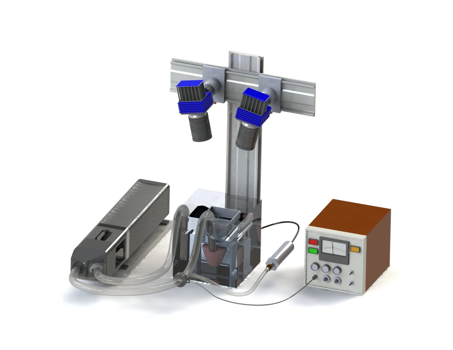
SolidWorks rendering of the experimental setup
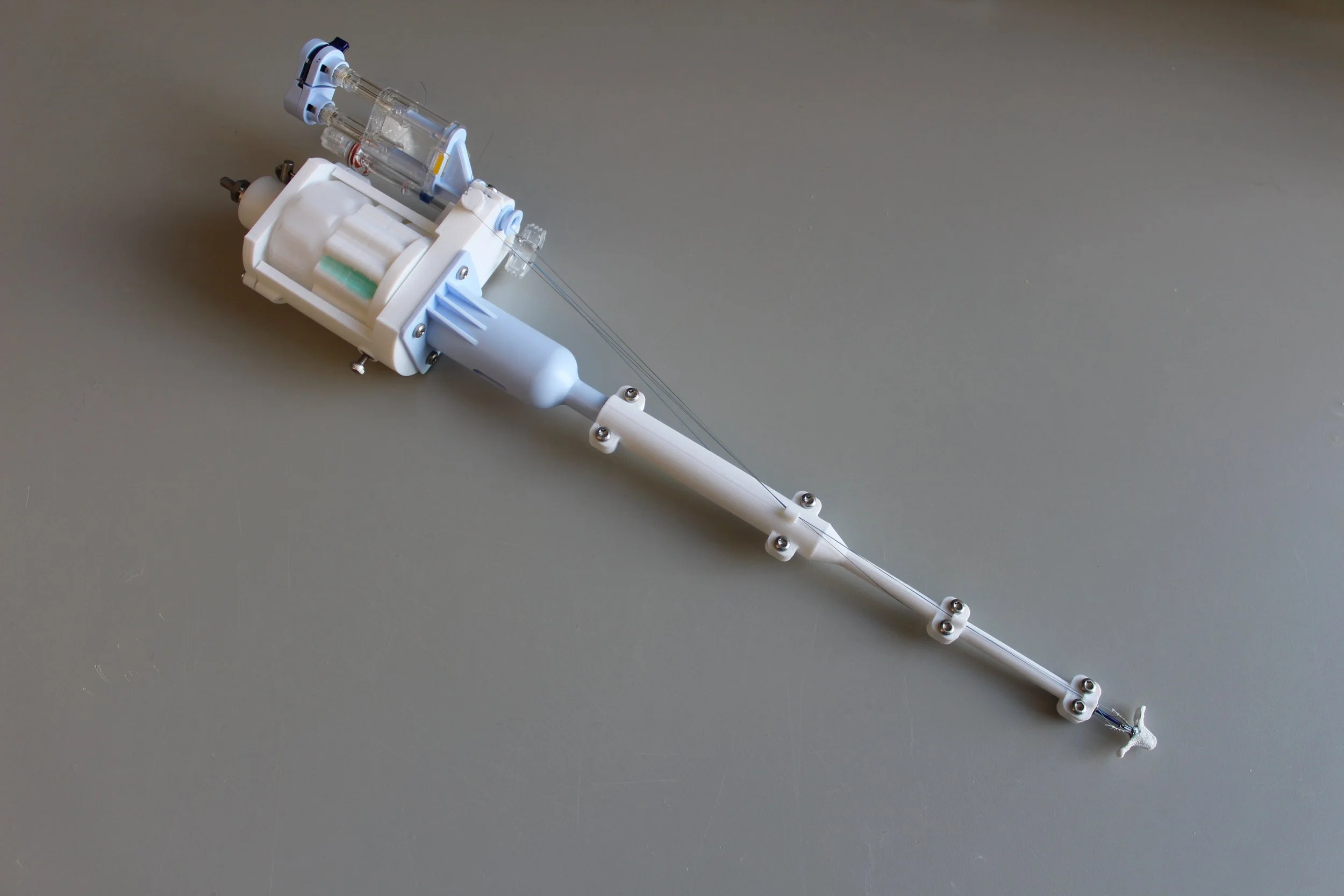
Custom 3D printed TriClip deployment device
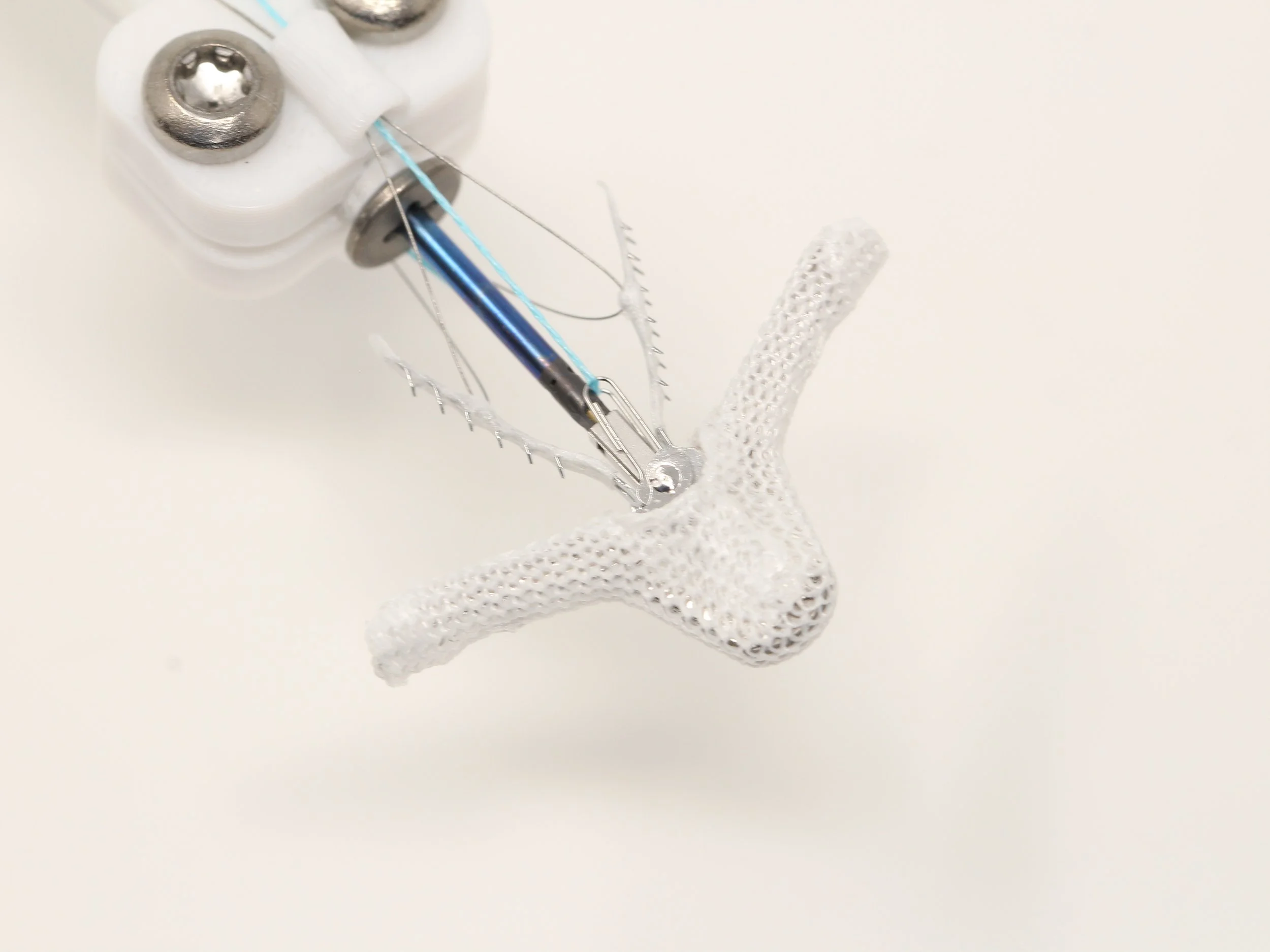
TriClip NTW
A sample dataset set representing forces measured at each point.
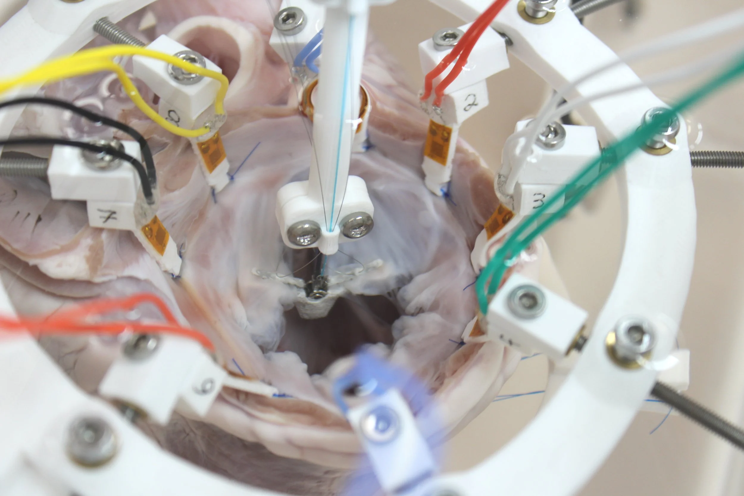
Performing TEER on a porcine heart
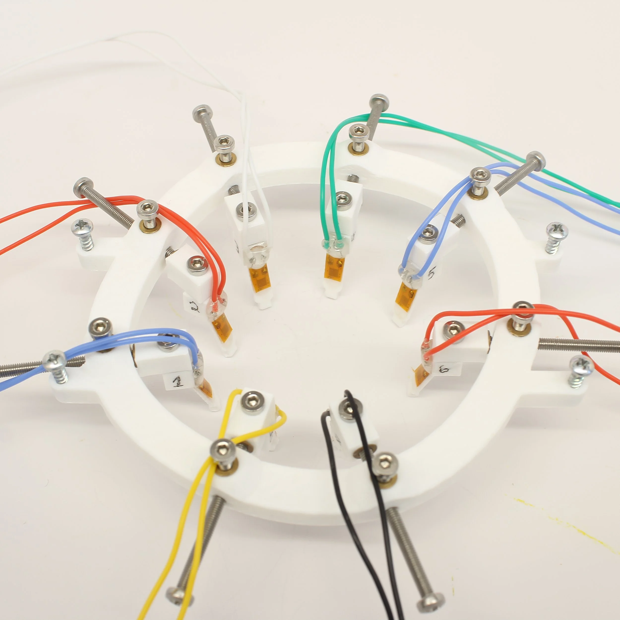
Heart mount with strain gauges
In-Vitro Evaluation of TEER Effects on Annular Forces with the TriClip
I developed an in-vitro platform to study how the transcatheter edge-to-edge repair (TEER) procedure alters annular forces around the tricuspid valve in hearts. The system integrates a dilatable heart mount with strain gauges bonded to 3D-printed structures acting as load cells at the annulus, enabling high-resolution measurement of local forces under physiologic loading. Explanted porcine hearts were mounted and pressurized via the pulmonary artery to simulate ventricular filling and systolic conditions.
Methodology
Disease Model - Mechanically dilated the annulus and applied controlled pressures to simulate tricuspid regurgitation.
TriClip Implantation - Developed a custom 3D-printed device for repeated placement of a normally single-use TriClip.
Data Acquisition - Recorded annular forces, ventricular pressure, and flow rate for each clip configuration.
Analysis - Processed and analyzed data in MATLAB and R.
Key Contributions
Designed and built a dilatable heart mount with an embedded load cell array.
Developed a reusable 3D-printed deployment system for controlled TEER interventions.
Conducted all testing, including sample preparation, TriClip implantation, and data collection.
Built the data analysis pipeline for quantitative assessment of annular forces.
Impact
This experimental design provides a reproducible in-vitro environment for directly assessing how TEER interventions affect annular mechanics, which may contribute to the remodeling of the right ventricular wall, a factor in the continued success of the procedure. The data collected from this experiment will enable the comparison of different TriClip placement locations, inform more effective surgical solutions, and support the design of more effective heart valve repair devices.