
SolidWorks Rendering of the experimental setup.
3D DIC of the tricuspid valve.
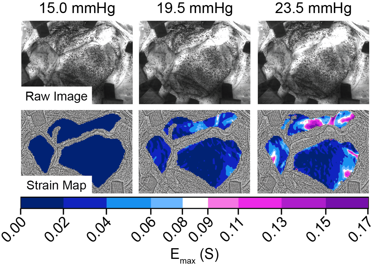
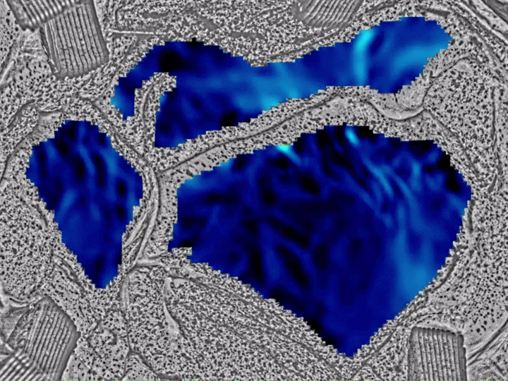
Sequential stain map from a sample data set.
In-Vitro 3D DIC Measurement of Tricuspid Valve Strain
I developed an in-vitro method to artificially disease and quantify 3D deformation and strain of porcine tricuspid valves using stereoscopic digital image correlation (DIC). The approach integrates a novel speckling protocol hydrated tissue, stereo imaging, and a full analysis pipeline from LaVision DaVis to MATLAB. This study has been submitted for publication and is under review.
Methodology
Speckle Patterning - Developed a novel method to apply speckle patterning to hydrated tricuspid leaflets.
Test Setup - Mounted valves in a submerged fixture under calibrated stereo cameras, with static water columns providing various pressures.
Disease Model - Applied controlled annular dilations with a custom, 3D printed annular dilation ring and pressurization through the pulmonary artery to induce regurgitation.
Imaging - Captured synchronized stereo image sequences during loading.
Data Processing - Reconstructed 3D point clouds in LaVision DaVis and exported to MATLAB for strain analysis and visualization.
Key Contributions
Designed and built the experimental setup.
Conducted testing, managing all aspects from sample preparation, test execution, and data analysis.
Built the full data analysis pipeline from generate 3D strain maps from image sequences.
Impact
This setup provides a novel and repeatable in-vitro method for quantifying 3D deformation and strain of tricuspid valves under varying physiologic conditions. This data will help improve understanding of valve mechanics to better inform the design of heart valve repair devices and current heart valve repair procedures.